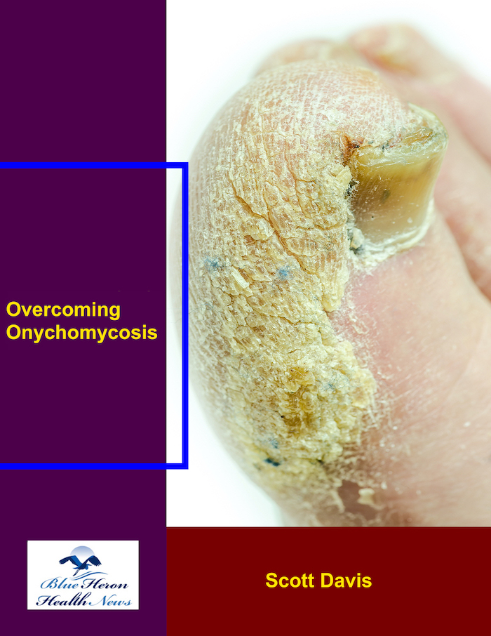
Overcoming Onychomycosis™ By Scott Davis If you want a natural and proven solution for onychomycosis, you should not look beyond Overcoming Onychomycosis. It is easy to follow and safe as well. You will not have to take drugs and chemicals. Yes, you will have to choose healthy foods to treat your nail fungus. You can notice the difference within a few days. Gradually, your nails will look and feel different. Also, you will not experience the same condition again!
What is the role of imaging studies in diagnosing onychomycosis?
Imaging studies are not often the first diagnostic strategy for onychomycosis, since the condition is usually diagnosed by a combination of clinical examination and microscopy of nail samples. Imaging studies may be an adjunctive measure in some cases, particularly when there is a need to evaluate more complicated or severe infections, or to rule out other ailments that may present in a similar manner to onychomycosis.
Here’s how imaging tests can be useful in diagnosing or treating onychomycosis:
1. Ultrasonography (Ultrasound)
Role: Ultrasonography of the nail could be used to quantify the nail thickness and estimate the extent of subungual involvement in onychomycosis. It is a non-painful technique that could provide a clearer picture of the nail and its anatomy.
How it Helps:
Distinguishing from other disorders: When there is uncertainty about whether an infection of the nail is fungal or if a pre-existing disorder (e.g., trauma or psoriasis) is present, ultrasound can be more informative.
Determining severity: Ultrasound can show the degree of thickening of the nail or disjunction from the nail bed, which is characteristic in onychomycosis.
2. X-ray Imaging
Role: X-rays are not used to diagnose onychomycosis on a regular basis, but in certain cases of involvement of the bone or suspected osteomyelitis, particularly in diabetic patients or those who are immunocompromised.
How it Helps:
Excluding bone infection: If there is severe inflammation and pain in the tissue around the nail, X-rays can detect osteomyelitis, which is a consequence of an unresolved fungal infection.
Nail deformities measurement: X-rays, in chronic or severe cases, can reveal deformed or traumatized nails that can be helpful in distinguishing onychomycosis from other causation of nail abnormality.
3. MRI (Magnetic Resonance Imaging)
Role: MRI is never used to make a direct onychomycosis diagnosis. It may be used where there is a need to assess deeper tissue involvement, especially in complicated or severe infections that have extended to adjacent structures.
How it Helps:
Detection of soft tissue involvement: If there is any suspicion that infection has spread into the surrounding soft tissues or into bone, then an MRI is helpful in determining how far the infection has spread. This is especially so for chronic infections or advanced cases where osteomyelitis is suspected or extensive tissue destruction is present.
Differentiation from other disorders: MRI is also used to rule out other disorders that might resemble onychomycosis, such as soft tissue tumors, or to assess abscesses or other complications.
4. Dermoscopy (Nail Surface Imaging)
Role: Dermoscopy is a trauma-free technique with the help of a specific magnifying tool (dermatoscope) to scan the surface of the nail and nail bed. The nail change can be viewed by the clinician closer than the naked eye with it, and it is helpful in differentiating fungal infection from other nail disease.
How it Helps:
Identifying characteristic features: In onychomycosis, dermoscopy can recognize features of white spots, yellow streaks, or coloration of the nail plate, all of which are classically associated with fungal infections.
Distinguishing from other conditions: It helps differentiate onychomycosis from other conditions like psoriasis, eczema, or nail trauma, which might show overlapping clinical features.
5. Role of Imaging in Chronic or Severe Cases
In severe, chronic, or complex onychomycosis, imaging procedures are able to provide valuable information upon which to make management decisions:
Assessment of therapy response: Imaging modalities like ultrasound can track response of the nail thickness or anatomy during treatment to enable practitioners to ascertain whether infection is being cleared or whether modification of therapy is necessary.
Detection of complications: In cases where the infection has caused complications like abscesses, soft tissue infections, or osteomyelitis, imaging can detect the degree of tissue damage as well as the necessity for further aggressive treatments.
6. Restrictions in Imaging
Even though imaging tests can prove helpful at times, they suffer from some drawbacks:
Not a diagnostic study for fungal infection: Imaging will not specifically identify fungal organisms or confirm onychomycosis. A microscopic examination of nail clippings or a fungus culture is needed to confirm the diagnosis.
Not typically required: In most patients with suspected onychomycosis, clinical examination and laboratory tests (e.g., KOH test, fungal cultures, or PCR testing) are sufficient to confirm the diagnosis, and imaging tests are not required.
Conclusion
Imaging tests, such as ultrasound or MRI, are not typically first-line diagnostics for onychomycosis but can be useful in establishing the extent of the infection, examining complications, or distinguishing between onychomycosis and other nail diseases in challenging cases. Dermoscopy is a valuable non-invasive imaging technique for observing changes in the nail and for distinguishing fungal infection from other nail conditions. A combination of clinical and laboratory examination in most cases is sufficient for definite diagnosis of onychomycosis.
Onychomycosis (nail fungal infection) in children is diagnosed using a combination of clinical evaluation, patient history, and investigations. The diagnostic process for onychomycosis in children is the same as in adults but requires special caution because nail infections in children sometimes imitate other diseases of nails in children. The following is the way onychomycosis is typically diagnosed in children:
1. Clinical Evaluation
Patient History: The prescriber will take an overall medical history, including the child’s symptoms, recent trauma, fungus exposure (e.g., communal showers or public swimming pools), and underlying illnesses (e.g., immunosuppression, eczema, or diabetes) that predispose them to infection with fungi.
Symptom Presentation: The doctor will look at the nails for signs that are characteristic of onychomycosis, including:
Discoloration: The nail may become yellow, white, or brown.
Thickening: The nail may thicken, become deformed, or brittle.
Brittle or Crumbly Nails: The infected nail is prone to crumbling or breaking easily.
Separation from Nail Bed (Onycholysis): The nail separates from the nail bed, allowing for a collection of debris or fungal material within the space.
Pain or Tenderness: The child may be tender or painful around the site of the infected nails, especially with a severe infection.
2. Differentiating from Other Nail Conditions
Differentiating onychomycosis from other nail pathology in children that is mimicking of fungal disease involves:
Nail trauma: Trauma to the nail can lead to changes in the nail appearance mimicking onychomycosis.
Psoriasis: Psoriatic children are susceptible to developing mimicking in nature nail changes of fungal infection, such as pitting, thickening, and discoloration.
Eczema: Atopic dermatitis or other eczema may lead to secondary changes in the nail like thickening or splitting.
Bacterial infections: Bacterial infections can also cause nail discoloration or separation in certain cases.
3. Laboratory Tests
Upon suspicion of onychomycosis, further tests are typically conducted to confirm the diagnosis and to identify the specific fungal pathogen:
Microscopic Test: Scrape a specimen from the affected nail or nail bed and inspect it under the microscope for signs of fungal hyphae (fungus filaments). It is a quite quick test to confirm the occurrence of a fungal infection.
Fungal Culture: A laboratory culture of the nail scrapings is performed to identify the causative fungus. It is utilized to check for dermatophytes (e.g., Trichophyton rubrum, Trichophyton mentagrophytes), yeasts (e.g., Candida species), or molds as the cause of the infection. This is more specific but takes longer (several days to weeks).
Dermatophyte Test Media (DTM): A more specific culture medium used for the growth of dermatophytes, which is also able to provide rapid results (3-7 days). DTM has the ability to differentiate between dermatophyte infections and other causes of nail alteration.
Polymerase Chain Reaction (PCR): Although not used in routine practice, PCR identifies fungal DNA in nail samples with more specific diagnosis and also identifies the individual species of fungi.
4. Imaging
X-rays: In some cases, if the infection is severe or is of the underlying structures of the nail, an X-ray may be used to assess the extent of nail and underlying bone damage.
5. Differential Diagnosis
Other causes of abnormal nails need to be ruled out, including:
Trauma: Trauma to the nail can cause analogous clinical changes to onychomycosis and hence the clinician should ask for a recent history of trauma or nail injury.
Psoriasis: In children, psoriasis can cause nail changes such as pitting, onycholysis, and nail thickening that are clinically similar to fungal infections. Careful search for other cutaneous manifestations of psoriasis (e.g., scaly plaques) will distinguish between the two.
Eczema: Atopic dermatitis and eczema can produce secondary nail changes, including thickening and splitting, which can mimic onychomycosis.
Bacterial Infections: Secondary bacterial infection can sometimes create nail changes simulating fungal infections, particularly in paronychia (paronychia is inflammation of the folds of skin next to the nail).
6. Special Considerations in Children
Immunocompromised Children: Immunocompromised children with immune deficiencies, diabetes, or those on immunosuppressive therapy (e.g., steroids) are at higher risk for fungal infections, including onychomycosis. It may be harder to diagnose in these children since they may have atypical presentations or more severe infections.
Toe vs. Fingernail Involvement: Onychomycosis is more commonly seen in the toenails due to prolonged exposure to humidity and the occlusive condition of shoes. Fingernails can be involved, especially in children with a history of trauma or fungal exposure.
Nail Appearance in Children: Nail appearance in children may differ from that of adults, and some fungal diseases may have mild symptoms or even local changes with other states simulated.
Conclusion
Diagnosis of onychomycosis in children is established on the basis of the complex of clinical presentation, patient’s history, and laboratory investigation (microscopic examination, fungal culture, etc.). The condition can generally be mimicked by other nail conditions, and thus the distinction between onychomycosis and others including trauma, psoriasis, and eczema has to be made. Fungal culture and microscopy are the critical diagnostic tests, while newer tools like PCR may offer specific identification of the fungal organism. Early treatment and diagnosis must be done in order to prevent sequelae like damage to the nail or secondary infection.
Overcoming Onychomycosis™ By Scott Davis If you want a natural and proven solution for onychomycosis, you should not look beyond Overcoming Onychomycosis. It is easy to follow and safe as well. You will not have to take drugs and chemicals. Yes, you will have to choose healthy foods to treat your nail fungus. You can notice the difference within a few days. Gradually, your nails will look and feel different. Also, you will not experience the same condition again!