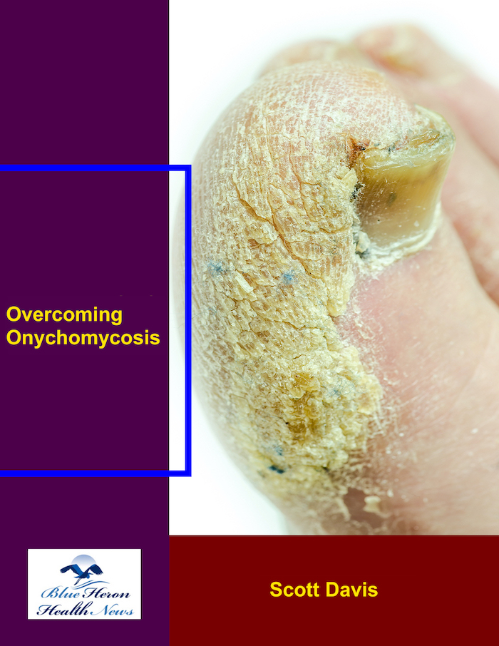
Overcoming Onychomycosis™ By Scott Davis If you want a natural and proven solution for onychomycosis, you should not look beyond Overcoming Onychomycosis. It is easy to follow and safe as well. You will not have to take drugs and chemicals. Yes, you will have to choose healthy foods to treat your nail fungus. You can notice the difference within a few days. Gradually, your nails will look and feel different. Also, you will not experience the same condition again!
What are the signs of onychomycosis under a microscope?
Signs of Onychomycosis Under a Microscope
Onychomycosis is a fungal infection of the nails, often caused by dermatophytes (fungi that infect skin, hair, and nails), yeasts, or molds. A microscopic examination of a nail sample is one of the most reliable ways to confirm the diagnosis. When a sample is taken from the infected nail and examined under a microscope, several characteristic signs of onychomycosis can be identified.
Here’s a breakdown of the key microscopic features that indicate onychomycosis:
1. Fungal Hyphae or Mycelia
- Hyphae are the branching filamentous structures that make up the body of fungi. In an infected nail, hyphae can be seen in the nail bed, nail plate, or in the nail debris.
- Dermatophytes (the most common causative agents) form long, thread-like structures that can be seen clearly under a microscope. The hyphae appear as thick, septate (divided by cross-walls), and branching filaments.
- Yeasts (e.g., Candida species) may also produce hyphal forms, but these are typically more rounded and less branched than dermatophyte hyphae.
2. Nail Plate Disruption
- The fungal infection often causes damage to the nail plate. Under the microscope, you may observe:
- Separation of the nail plate from the nail bed (onycholysis).
- Thickening or splitting of the nail plate, which is often seen with more chronic infections.
3. Spore Formation
- Conidia or spores are often produced by the fungi and can be seen under the microscope as small, round or oval structures. These are typically seen in:
- Dermatophyte infections: Conidia appear as round or oval structures in chains or clusters.
- Yeast infections: Candida species, for example, produce oval to round spores that may be single or in groups.
4. Nail Debris
- Infected nails may have discolored, crumbled debris that contains fungal elements. Under a microscope, this debris may contain fungal hyphae and spores. The debris is often keratinized material that has accumulated from the damaged nail plate.
5. Inflammation and Immune Response
- In some cases, the immune response to the infection may be visible in the form of:
- Inflammatory cells (such as neutrophils or lymphocytes) may be observed around the infected areas of the nail. This is a sign of the body trying to fight off the infection.
- The presence of these cells can also help distinguish between fungal infection and other nail conditions like psoriasis or eczema.
6. Fungal Filaments in Nail Bed or Matrix
- In more advanced infections, fungal elements may be seen in deeper parts of the nail, such as the nail bed or nail matrix. This is typically seen in chronic or severe cases of onychomycosis and helps confirm the extent of the infection.
7. Type of Fungus
Under the microscope, the specific type of fungus can often be identified, which helps with determining the appropriate treatment:
- Dermatophytes (e.g., Trichophyton, Epidermophyton, Microsporum) are the most common cause of onychomycosis and have distinctive hyphal forms.
- Yeasts like Candida can cause onychomycosis and often form larger, budding cells that look different from dermatophyte hyphae.
- Molds (e.g., Aspergillus, Fusarium) may show more branching and irregular hyphal growth, often appearing darker under the microscope.
Conclusion
When onychomycosis is suspected, microscopic examination of a nail sample can confirm the diagnosis by revealing fungal elements like hyphae, spores, and nail plate disruption. The specific features seen under the microscope can help determine the type of fungus responsible for the infection and guide appropriate treatment decisions.
Would you like to explore how onychomycosis is treated or more about fungal culture methods for confirming diagnosis?
Overcoming Onychomycosis™ By Scott Davis If you want a natural and proven solution for onychomycosis, you should not look beyond Overcoming Onychomycosis. It is easy to follow and safe as well. You will not have to take drugs and chemicals. Yes, you will have to choose healthy foods to treat your nail fungus. You can notice the difference within a few days. Gradually, your nails will look and feel different. Also, you will not experience the same condition again!